 Temple Grandin, PhD, is likely the most well-known, living autistic individual—both nationally and internationally. As Distinguished Professor of Animal Science at Colorado State University, she is equally famous as an astute observer and researcher of animal science and behavior, especially in designing humane livestock-handling systems. As a stellar scientist and educator driven by scientific inquisitiveness, Dr. Grandin extended her writings and research toward an improved understanding of autism, including how her own journey and insights could provide testable hypotheses about its neurobiological underpinnings.
Growing up in the 1940s through the 1960s, Dr. Grandin battled misperceptions about autism, which sparked her first writings about coping strategies (Grandin, 1983). She then asked more fundamental scientific questions about the neurobiology of autism, which moved toward personal speculations about brain–behavior relationships and the autism spectrum (Grandin, 2009a, 2009b; Ratey et al., 1992). In these later publications, Dr. Grandin wrote about how she processes sounds and visual stimuli, speculating on how differences in brain organization and connectivity may relate to autistic features. When lecturing about autism, Dr. Grandin routinely discussed how she “thinks in pictures, not words.”
In this personal quest, knowing how important a unique case study can be in medicine and psychology, Dr. Grandin underwent magnetic resonance imaging (MRI) of her brain, working with investigators at the University of Utah, the University of Wisconsin–Madison (UW), and Brigham Young University (BYU), who had formed the Interdisciplinary Science to Learn about Autism collaborative research program. These extensive MRI studies involved both structural and functional MRI, which included state-of-the-art quantitative neuroimaging analyses as well as a battery of neuropsychological tests. Current advances in quantitative neuroimaging, combined with neuropsychological assessment findings, now permit a more comprehensive understanding of brain–behavior relations by directly examining brain MRIs.
In a case study published in the Journal of Pediatric Neuropsychology, Erin David Bigler and colleagues summarize the initial neuroimaging investigations involving brain morphometrics and Dr. Grandin’s functional neuroimaging. These studies integrated her brain-imaging data with her neurocognitive profile, derived from standard methods of neuropsychological assessment. This article is the first of a series showing how advanced neuroimaging can provide unique insights potentially relevant to understanding variation in autistic adults.
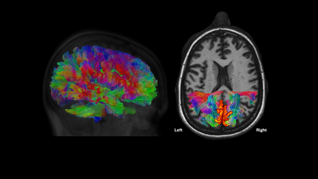
The figure below shows one technique: diffusion tensor imaging. The lateral image on the left reflects whole-brain white matter tractography, depicting the complexity of how tracts project to different brain regions. The image on the right demonstrates how more detailed analyses of specific tracts can be extracted, providing insights into the relationship between brain structure and cognitive function.
Dr. Grandin’s brain. (Left) Lateral view of the whole brain showing white-matter tracts. The coloration is significant. Green defines tracts projecting in the anterior–posterior plane; blue, vertically oriented tracts; and red, lateral projecting fiber tracts. (Right) Axial view with selected tracts in a region of the posterior corpus callosum involving projections with the precuneus region of each hemisphere. Note the asymmetry of the lateral ventricle (dark region in the center of the structural MRI), which is discussed in the case study.