
At the Mayo Clinic in Jacksonville, Florida, surgeons opened a patient’s skull, preparing to remove a tumor located in a high-risk area of his brain. Then, they woke the patient up. The neurosurgeon used a probe to stimulate the brain tissue, mimicking what would happen if that piece of tissue was sliced through or surgically removed. Meanwhile, clinical neuropsychologist David Sabsevitz, PhD, ABPP, tested the patient’s cognitive functions. Asked to name a flesh-eating fish, the patient responded “pirannel” and “pirannus” but was unable to remember the word “piranha.” Asked to repeat the nonsense word “sakanting,” he could only reply with “sakanking” and “sakankus.”
Though subtle, those changes indicated that the probe was encroaching on important language areas of the brain. Sabsevitz’s tests helped the surgeon avoid those critical areas and find a safe corridor through which to access and remove the tumor—without causing a significant language deficit.
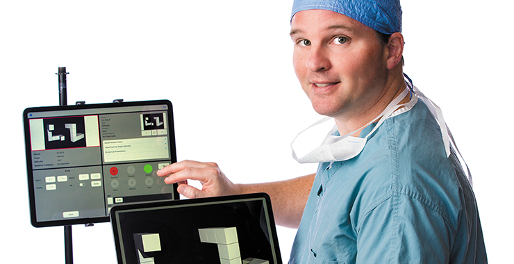
Such precision is possible because of a device called the NeuroMapper, a tabletbased testing platform that helps neuropsychologists and other clinicians map and monitor critical brain functions during surgery to treat brain tumors and epilepsy. Sabsevitz developed the system in 2016 while at the Medical College of Wisconsin, in a joint effort with computer science students at the University of Wisconsin–Milwaukee. Now a senior associate consultant at the Mayo Clinic in Jacksonville, he has used the platform with more than 200 surgical patients and counting and continues to fine-tune it. The Monitor spoke with Sabsevitz about the ways that the NeuroMapper is helping patients and improving our understanding of the brain.
Why do you map brains during surgery?
When operating on patients with brain tumors or epilepsy, surgeons may be working close to important areas such as those for language, sensory or motor functions. Before patients even enter the operating room (OR), the surgical team does structural and functional brain imaging to map the brain anatomy and the connections between brain areas. We then take those data, as well as the data I get from evaluating patients with traditional paper and pencil tests, to develop a game plan before we go into the OR. Then we wake the patient up for part of the surgery so we can map important functions and avoid those areas. While there are similarities in how brains are organized, it’s remarkable how much variability there is between individuals. Add in a disease like a brain tumor or epilepsy, and you can have even more differences in brain organization. If we did these surgeries while patients were asleep, we’d be shooting in the dark.
How does NeuroMapper improve on the process?
Practices for brain mapping are incredibly variable across hospitals. Many sites will talk to the patient casually, looking for gross changes in conversation skills. Many show patients pictures and ask them to name the objects as a way of testing language ability. But very few sites use multidimensional mapping, testing a variety of functions using conceptually sophisticated measures. My colleagues and I wanted to increase the sensitivity of these tests to identify any changes in functioning as early as possible. With the NeuroMapper, the patient views a tablet mounted on a bar attached to the operating room bed. I have a second tablet that faces me, and I can select what tests I want the patient to see, and track the results. It’s obvious if a patient can no longer name a picture that they were able to name before surgery. But with NeuroMapper, we can pick up on subtle changes, such as changes in reaction time. Maybe they were able to name a rhinoceros in three seconds, but now it takes them six seconds. That tells us the surgeon might be getting too close to language areas, and we should proceed with more caution. In the past, we couldn’t measure those subtle changes very well.
What impact is it having on patients?
We’re operating on patients that we might not have operated on before, such as patients with tumors close to brain regions critical for language or other cognitive functions. Without the proper methods to map and monitor them, such surgeries were often too risky. Neurosurgeons are also able to be more aggressive in removing tumors, because they’re getting constant feedback about the impact of surgery as the surgery is progressing. They can keep going as long as they can see the patients are doing well.
At the same time, it’s helping us learn more about the brain. We’re developing new types of neuropsychological tests that allow us to map parts of the brain we weren’t mapping in the past. I think it will lead to better surgical outcomes, but I also see this as a tool to facilitate our understanding of brain functioning.
What kinds of things are you finding?
We’re learning more about the nondominant hemisphere, for example. That’s the nonlanguage hemisphere, and for most people it’s the right side of the brain. In the past, surgeons thought operating on the nondominant side wasn’t as risky, so most of these surgeries were done while patients were asleep. But there are data suggesting cognitive risk in these cases. NeuroMapper gives us novel ways to try to map these areas. Also, for example, brain regions involved in attention are distributed throughout the right hemisphere. If you damage parts of that network, you can get what’s known as neglect. You can see what’s in front of you, but your brain doesn’t pay attention to what’s happening on the left side. You wouldn’t see things on your left side, and you’d bump into them. If you were driving, you might veer to the left. We’ve developed a number of measures that allow us to map and monitor these attentional networks during surgery. We’ve been able to pick up early signs of neglect to spare patients more significant neglect after the operation.
What are your plans for NeuroMapper going forward?
It’s now being used in about 20 academic medical centers across the United States, including here at the Mayo Clinic. We don’t charge for it, but there is a selection process we use to decide who to make it available to. That’s because one of our goals is to create a network for collaborative research with academic institutions across the country. I look at this as an opportunity to improve standardization and research. If we have a tool that’s standardized, we can do research across institutions and collect data much more efficiently than we’ve been able to do.
NeuroMapper has been through several iterations since we started using it in 2016. We’re constantly developing new tasks to help us measure areas of the brain we don’t understand very well. We’re also collecting outcome data, following patients before and after surgery to see what effect this is having. We learn new things every day. The brain is fascinating, and this is a really exciting space to work in. But getting a patient through a really difficult surgery and seeing them function well afterward—that’s what this is all about.