
Some one in five patients drop out of mental health-care treatment, often because their first treatment didn't work. Is there a way to predict what will?
A growing number of psychologists and other scientists are using MRI, fMRI and PET on a quest to find out. Although it's still in the very early stages, their research suggests that clinicians may one day be able to match patients to effective treatments and ease symptoms faster by using information from brain imaging, along with other biomarkers such as DNA and hormone levels.
"It's possible that within our lifetimes you will go to your doctor's office, give blood, and if it's not prohibitively expensive, get an fMRI," says Gabriel Dichter, PhD, an associate professor of psychiatry at the University of North Carolina. "We accept it as part of standard care of neurology and cancer care, so why not for mental health?"
Finding best responders
Current treatments for mood and anxiety disorders are trial and error. Studies show that only about half of patients respond to their first course of treatment, whether it's psychotherapy or medication, or both. Pinpointing what works can take months, which is "just not acceptable for people potentially having suicidal thoughts," Dichter says.
Further, symptoms can also differ widely even within the same disease. "Two people can be diagnosed with major depression and not have a single symptom in common, so you're setting yourself up to fail to think the same treatment will necessarily be effective for those two patients," he says.
Researchers are trying to predict the outcomes of different treatments by examining patients' brains pre- and post-therapy. Among them are Dichter and Moria J. Smoski, PhD, of Duke University and colleagues. Using resting-state fMRI, they looked at the brains of 23 people with depression before behavioral activation therapy, which has been shown to affect how the brain responds to rewards. The team found that people whose depression improved most had greater brain network connectivity between the anterior insular cortex—a region involved in assigning importance to events—and the middle temporal gyrus, which plays a role in the subjective experience of emotion (Neuropsychopharmacology, 2016).
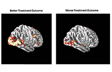
 Examining patterns of brain connectivity also helped researchers predict how well people did with cognitive behavioral therapy (CBT) for social anxiety disorder. Using scans of people's brains in a resting state—awake, but not focused on a particular task—Susan Whitfield-Gabrieli, PhD, of the Massachusetts Institute of Technology (MIT), and colleagues found that differences in both brain structure and neural connectivity among different regions predicted how well CBT reduced people's symptoms. Clinician estimates of treatment outcome using a behavioral assessment tool were accurate only 12 percent of the time, but adding information from imaging improved estimates of treatment success fivefold (Molecular Psychiatry, 2015).
Seeing how people's brains perform when they're doing complex tasks may also shed light on how they will respond to therapy. So far, research suggests that people with social anxiety pay excessive attention to potential threats, which undermines their ability to process neutral information. Heide Klumpp, PhD, of the University of Illinois at Chicago, scanned the brains of 32 people with social anxiety as they performed an emotional interference task, which asked them to identify certain letters behind which occasionally lurked pictures of angry faces. She found that participants who struggled most to avoid being distracted by the threatening stimuli—signaled by more activity in their dorsal anterior cingulate cortex—had the most improvement in their social anxiety symptoms when treated with CBT (Social Cognitive and Affective Neuroscience, 2016).
Those who had greater reactivity to the angry faces also showed less connectivity with certain frontal areas of the brain, a deficit that CBT may improve. So they had the most to gain, Klumpp says, adding, "Maybe how your brain is wired before a certain therapy speaks to what you're going to get out of it."
Measuring brain connectivity patterns with resting-state fMRI may predict who will relapse after receiving CBT for obsessive-compulsive disorder, according to a study by Jamie Feusner, MD, of the UCLA Semel Institute for Neuroscience and Human Behavior, and colleagues (Frontiers in Psychiatry, 2015). Patients with denser local brain connections that confer greater efficiency—known as small-worldness—were more likely to have a return of symptoms after CBT.
"The brains of people with OCD with high small-worldness may be like having a really fast Internet connection and a sophisticated search engine—they can zip from one thought to the next so quickly and pull up information so readily that it allows them to easily get deeper and deeper into obsessional thinking," Feusner says.
Drugs versus therapy
Research has also compared the brain profiles of people who respond to antidepressants versus psychotherapy. A groundbreaking study by Helen Mayberg, MD, of Emory University, and colleagues, measured brain glucose activity using PET. Results showed that less resting-state activity in the insula predicted that a person would be more likely to respond to CBT versus an SSRI, while more activity in the insula was associated with the opposite (JAMA Psychiatry, 2013).
But a larger question is whether it's realistic to have people routinely undergo brain scans to target treatment. "The worry I have is, from a pure cost basis, will it make sense to do an MRI or just give people a few trials of medicine? Or can we use a cheaper cognitive test as a surrogate marker of brain activity?" asks Warren Taylor, MD, of Vanderbilt University, who is leading a five-year study funded by the National Institutes of Health using MRI to measure vascular issues that might affect brain structure and connectivity in older adults.
While imaging is still expensive, new technology may improve its value. For example, widely available and portable EEG machines can measure people's real-time brain activity between scans. And it's possible that MRIs may one day be read automatically, making it easier to see relative brain differences among different people and to process results faster.
Computer models will also help to help predict treatment outcomes by translating the now massive amounts of brain scan data and information on DNA sampling, as well as the many variables that can contribute to mental illness, including childhood trauma, poverty and sleep quality.
Though some rate of adoption seems inevitable, it's too soon to say when predictive imaging will be common, researchers say, and it will be easier to implement in integrated practices. Still, it's likely to find its way to clinical use faster than new treatments, says John Gabrieli, PhD, a neuroscientist at MIT.
"Everybody wishes for dramatically different treatments, but very little there has paid off," he says. "The choice of treatment is fairly haphazard these days, so for the foreseeable future, it would be progress if you could just steer a patient toward a treatment that's likely to work for him or her."